Friday, February 26, 2016

Third Week!

Hi everyone!

This was my third week at SRP and I have started to get a clearer understanding of where my project is headed and how I'm going to get there.  To start off, I spent this week with less time with my mentors and more time with people behind the scenes. I shadowed MRI technicians, who are the people that actually operate the MRI to scan whatever area needs to be scanned and learned that they are the ones to get the first look at the MRI scans. Most importantly in my third week at Mayo I learned that unlike what they show on Grey's Anatomy, these people are not surgeons. Instead, they are responsible for preparing the patient for the procedure and properly positioning them in the scanner. By shadowing the tech's, I learned that protocols are a combination of MRI sequences to optimally assess a particular region of the body. If this is a little confusing try to think of someone taking a test. Ideally, they would want to get a 100% but to do so, they would need to pick all the correct answers for every question. Just like this, when running a protocol, technicians must set the "correct" settings to effectively scan the patient to get the needed images. Radiologists are the ones who make the protocol, based on the patient's condition, and the MRI tech's are the ones who run the MRI protocol on the MRI.

While shadowing the technicians, I got an opportunity to run one of the scans myself and I even got up close and personal with the MRI machine! When I went inside the room with the MRI I was reminded 100 times not to bring in my phone or credit card because the magnet is so strong that it erases the credit card and renders it useless. For some reason I had been under the impression that every time the MRI machine was not in use, the technicians turned it off somehow and turned it on again every time it was needed. But the closer I got to the machine, I could feel how strong the magnet was because the magnetic part of my lanyard containing my badge was floating mid-air towards the MRI and I had to fight against the pull of the magnet to stay in one spot! While that experience was very exhilarating, actually running one of the scans was very interesting too. Although the technicians told me exactly what to input for the protocols and all I really did was click the buttons they instructed me to, I learned that running the scans requires a lot of prior knowledge about over a thousand protocols.

This week was much more hand-on than before and an unforgettable experience, especially with the MRI magnet pulling me towards it! Hopefully next week will be just as eventful!

Friday, February 19, 2016

Second Week!


Hello everyone! In my second week of SRP, I spent a lot of it researching the technical and programming side of an MRI and the physics behind how it functions.


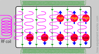
As you know, humans are mostly made out of water molecules and these molecules are found throughout our bodies in different ways. MRI machines can’t detect the electrons of hydrogen or the oxygen atoms; rather they “see” only the nucleus of the hydrogen atoms contained in the water molecule. These machines use properties of the hydrogen nuclei to produce MRI scans. As you might recall from honors chemistry, atoms have a property called “spin” and the spin of the hydrogen nuclei can be oriented in certain ways. The magnetic field of the MRI makes the spins of most of the hydrogen nuclei line up along the magnetic field. Few hydrogen nuclei however will have spins opposite to the magnetic field and these nuclei will be “high energy nuclei” while the others will have “low energy nuclei”. The green arrows in the picture below symbolize the magnetic field and the blue arrows show the spin.


When the low energy hydrogen nuclei are bombarded or “irritated” with energy, they respond in a way that allows us create a scan. But how does their response create a scan? Well every MRI machine has a coil of wire that produces enough energy to irritate the low energy hydrogen nuclei. When a current is applied to that coil, which is often called a RF coil, energy is produced in the form of a rapidly changing magnetic field, shown by the pink waves in the diagram.


This energy that is emitted  from the RF coil is the main building block of the MRI. The low energy nuclei absorb this RF energy and change their spin direction and become high energy nuclei.   


After some time, the RF energy is stopped and the recently turned “high energy nuclei” go back to their previous low energy state and as they do this, they start releasing the energy that was given to them. They release this energy in the form of waves and these waves are collected by “receiver coils” which is the blue coil in the animation. The receiver coils convert the energy waves into an electrical current signal and through this, the MRI machine can perceive the amount and concentration of hydrogen nuclei in the body.


Here you can see how an actual MRI scanner looks with all the pieces of the animations. 


While the physics behind an MRI is certainly confusing, I hope I cleared up some questions. I hope to get a more concise perceptive next week at my internship. Hope you all stick around till then!

Friday, February 12, 2016

First Day!


02/12/16

Hi everyone and welcome to my second blog post!


This week I didn’t get to spend too much time on-site at Mayo since my first official day was today, Friday. I focused some of my time this week on independent research. I attempted to decode several images Dr. Panda, my co-mentor, had sent to me prior to the start of my project to help with my proposal. I wasn’t able to decipher much at first, but now looking at it again after I went over the basics of a MRI machine today, it makes a bit more sense.


On my first day, Dr. Panda introduced me to many doctors including my other co-mentor Dr Silva. I toured the Radiology department in the Mayo Clinic Phoenix location and learned that they have seven MRI’s up and running almost all the time. We spent most of the time discussing how I would carry out my project and what they could do to assist. Most patients getting a MRI are either getting cranial scans or spinal scans but the patient scans that I will be looking at will be from the prostate and possibly the liver or pancreas. Dr. Panda and Dr. Silva explained to me that a MRI scan is composed of many series and a series is a picture that gives a different bit of information about what is being scanned. Each series comprises of different sharpness, resolution, etc. that tells us something new. Sometimes when people aren’t able to sit through a full MRI scan, doctors are left with only a few series or pictures and not the complete scan. Some series give more valuable information necessary for a diagnosis than others and with the help of Dr. Panda and Dr. Silva, I will be using a trial of prostate scans to determine which series are most important.

After deciding that using prostate scans will be most effective for the research project, Dr. Panda and I went to the Scottsdale campus of Mayo Clinic where I sat in on a meeting. I met with other doctors such as Dr. Min and Dr. James, both of whom I will be interacting with more later in my project. In the meeting they discussed, how to utilize the MRI machines so that there an optimal number of exams performed per hour. After the meeting, Dr. Panda and I returned to the Phoenix campus and finalized our schedule. Due to his and Dr. Silva’s unpredictable hours, the times when I will go in and shadow will be determined on a weekly basis. From my experiences today, I am looking forward to going in sometimes next week to meet with the rest of the team who I will be working with and to finally get started on collecting data for my project!