Hello everyone! In my second week of SRP, I spent a lot of it researching the technical and programming side of an MRI and the physics behind how it functions.
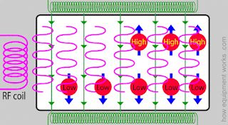
As you know, humans are mostly made out of water molecules and these molecules are found throughout our bodies in different ways. MRI machines can’t detect the electrons of hydrogen or the oxygen atoms; rather they “see” only the nucleus of the hydrogen atoms contained in the water molecule. These machines use properties of the hydrogen nuclei to produce MRI scans. As you might recall from honors chemistry, atoms have a property called “spin” and the spin of the hydrogen nuclei can be oriented in certain ways. The magnetic field of the MRI makes the spins of most of the hydrogen nuclei line up along the magnetic field. Few hydrogen nuclei however will have spins opposite to the magnetic field and these nuclei will be “high energy nuclei” while the others will have “low energy nuclei”. The green arrows in the picture below symbolize the magnetic field and the blue arrows show the spin.
When the low energy hydrogen nuclei are bombarded or “irritated” with energy, they respond in a way that allows us create a scan. But how does their response create a scan? Well every MRI machine has a coil of wire that produces enough energy to irritate the low energy hydrogen nuclei. When a current is applied to that coil, which is often called a RF coil, energy is produced in the form of a rapidly changing magnetic field, shown by the pink waves in the diagram.
This energy that is emitted from the RF coil is the main building block of the MRI. The low energy nuclei absorb this RF energy and change their spin direction and become high energy nuclei.
After some time, the RF energy is stopped and the recently turned “high energy nuclei” go back to their previous low energy state and as they do this, they start releasing the energy that was given to them. They release this energy in the form of waves and these waves are collected by “receiver coils” which is the blue coil in the animation. The receiver coils convert the energy waves into an electrical current signal and through this, the MRI machine can perceive the amount and concentration of hydrogen nuclei in the body.
Here you can see how an actual MRI scanner looks with all the pieces of the animations.
While the physics behind an MRI is certainly confusing, I hope I cleared up some questions. I hope to get a more concise perceptive next week at my internship. Hope you all stick around till then!


Wow this is super interesting! I never knew that MRI machines fluctuate between high and low energy phases!
ReplyDeleteFantastic! Now I have a clearer picture (no pun intended) of how an MRI works. Great job, Sparshee!
ReplyDeleteThese diagrams have been incredibly helpful in understanding MRIs, thanks for posting them!
ReplyDeleteWhat have you found most enjoyable about your internship so far?
Hi Ms. Conner! I've definitely enjoyed meeting a wide variety of doctors in different professions! It's been eye-opening and I never knew there were so many positions in the Radiology department.
DeleteWoah this is super neat! Did you find this out independently or did your mentor teach you this stuff?
ReplyDeleteI found the basics of how the machine works from my mentor but I had to research it more in depth to really figure out how an MRI machine is operated!
DeleteI agree with Ms. Conner; the diagrams helped me a lot too! Thanks for another didactic post where I learned something new!
ReplyDelete