Friday, April 22, 2016

Here's a link to my final SRP presentation!

https://docs.google.com/presentation/d/11B8cmHTxLdjUaVQ4_wT11p9DV3Bss-5hqtYo6gtyMOE/edit#slide=id.g129da60d9f_1_10

Enjoy!


Monday, April 18, 2016

Week Nine!

Hi all!

In my ninth week of my internship I decided that I would be extending my time at Mayo Clinic and doing research there outside the scope of SRP! My research questions delves into which scans are the most important in diagnosing prostate cancer and the research I will be doing outside of SRP will further explore the potential of these scans. My mentors and I will determine whether whether a shortened protocol with only these important scans is just as effective as the full length 45 minute protocol that is currently in use. Since the research I will be doing there still relates to my project, I will use any findings to support my SRP presentation.

So in the past week I did more personal research than I spent time at the clinic. My mentors assigned me to look at past articles on the effectiveness of a shortened protocol since we don’t want to be conducting research that’s already been done. As I’ve mentioned in my last blog post, the three most important scans are the T2, diffusion, and perfusion scans. Some of the articles I found compared the T2 and the diffusion with the T2 with the combined diffusion and T2 being more effective.

A phrase I saw come up often in all the reports I read was ROC curve and initially, naive me thought it was a radiology term. Coincidentally when I went to meet with Dr. Panda he gave me an article to read about Receiver Operating Characteristic (ROC) curves and how they relate to medicine. ROC curves basically weigh the pros and cons of a test such as its accuracy, amount of type 1 and 2 errors, etc. and essentially determines how good of a test it actually is. For example, breast cancer occurs around 3 out of 1000 people. A breast imager looking at 1000 cases could call all of them negative without ever really seeing them and have an accuracy of 99.7%. However this isn’t a reliable way to find the quality of a test. An ROC curve uses the number of true positives and true negatives, as well as false positives and false negatives and other variables to determine the proficiency of a test. Therefore, the larger the area under the ROC curve is, the “better” a test is.

Thank you all for reading and I hope to see you next week for my final blog post!

Friday, April 8, 2016

Hi everyone!


As I said in my last post, the brightness and darkness of certain areas on the MRI scans indicates the existence of prostate cancer and this week I found out which kinds of scans and which area of the scans give the most vital information. This came pretty close to answering a major part of my research question so I was very relieved! If you recall, my research question was “When a patient can’t complete an MRI scan, can a ‘partial’ exam be used as a substitute?” And with the information I learned this week, I gathered what this ‘partial’ exam would mainly constitute of.


In a prostate protocol the three most important scans are the t2, diffusion, and perfusion. What does that mean? Good question.


I’ve mentioned t2 before and as a recap it’s an imaging technique where liquid will appear bright and any fat will appear dark. Diffusion is an imaging technique that uses the random movement of water molecules to generate contrast in MR images. It maps the diffusion process of different molecules, mainly water, in biological tissues. Since molecular diffusion reflects interactions with many obstacles such as other macromolecules, fibers and membranes, and an MRI creates scans using these molecular diffusion patterns to reveal details about tissue architecture. Perfusion MRI is based on the analysis of MRI images after the peripheral injection of a contrast agent. Perfusion itself refers to the process of a body delivering blood to a capillary bed in its capillary tissue and perfusion imaging exploits vascular abnormalities and altered flow dynamics that lead to changes in blood volume and flow.

I will explore how the appearance of the prostate in these three images indicates cancer. Until next time!

Friday, March 25, 2016

Week 6!

Hi everyone and welcome to week 6 of SRP!


This week I spent more of my time with Dr. Silva interpreting images as opposed to sitting with the MR techs and watching the images being produced like I have been the last few weeks. I started of this week in the reading room watching the doctors go through scans and recording their observations about them. I was with them for about an hour before I realized they were interpreting CT scans and not MRI scans. CT’s are different from MRI’s because they take about a minute to scan the patient whereas MRI’s take around 30-45 minutes. Not only are they faster but they are much cheaper and more comfortable for the patient. However, CT scans are mostly useful for bone injuries while MRI’s are useful for soft tissue evaluation such as ligament and tendon injuries, brain tumors, and, pertinent to my project, prostate cancer.


In the CT scans I saw the doctors diagnosing, they were looking specifically for kidney stones. The stones appeared bright white on the scans since calcium shows up white. This is also why bone is very white in the CT scan below.  


The tiny white circle indicates the kidney stone and many times the stones are so small they are not detected by the doctors.


After spending some time interpreting the CT scans, I moved on to researching more about the prostate itself since the answer to my research question is backed up with evidence regarding the prostate and prostate cancer. I learned that there are two main parts to the prostate that shows up on MR scans.


The central gland and peripheral zone are the two parts that are examined to see whether or not the patients has prostate cancer. As suggested by the name, the peripheral zone surrounds the central gland and is seen as bright white on the picture above. The cancer is mostly in the peripheral zone which is why the zone is so enlarged in the picture. On a cancer-free patient, a prostate MRI would look like in the image below.


Dr. Silva taught me that the appearance of the peripheral zone and central gland on certain MRI series and the brightness or darkness of certain areas is a big indicator of whether or the patient has cancer. However, sometimes there are false positives and in the case of false positives, doctors must rely on some scans more than other. I will go into this more next week and I hope to see you then!

Saturday, March 19, 2016

Week Five!

Hi everyone!

This week on the fifth week of my internship, I explored the differences between MRI machines themselves. The Scottsdale campus of Mayo Clinic has around four MRI’s with a power of 1.5 Tesla while the Phoenix campus has around seven 1.5 Tesla MRI’s and a few more MRI’s with powers of 3 Tesla. Without going too much into the physics of it, a Tesla is the standard unit of magnetic flux density and is equivalent to one kilogram per second squared per ampere.

But what’s the distinction between the 1.5T vs. 3T? Is a 3T MRI twice as good as a 1.5T MRI? Sure, as far as the magnetic field strength is concerned. Price-wise a 3T MRI is also usually double the cost of a 1.5T. However, a 3T MRI is not necessarily twice as adept as the 1.5T. There are many drawbacks to a 3T even if its stronger magnetic field will allow for better image clarity. For example the scan times for a 3T are much longer and as my project explores, the longer the time the scans take, the more likely patients are to quit the scans unfinished, causing an inaccurate diagnosis or other problems.

A 1.5T MRI is adequate for the majority of scan that are done on all MRI’s today so there isn’t much of a necessity for the 3T’s. But since the stronger magnetic field of the 3T increases the signal available which is what actually creates the image, the definition of the scans produced on the 3T are actually better. However, they are better at a cost and that cost is the increased scan times.

In every MRI scan there is always image noise. While image noise is not actually a sound, it interferes with the signals given out to create images and actually comes from the patient’s body and other electronics inherent in the MRI. Ideally, there would be no noise present but this isn’t possible so the goal is to have more signal in making the image than noise which can affect the quality of the image. The 3T and 1.5T both achieve this but in different ways. A 3T MRI gives out more signal or more molecules that are resonating in the greater magnetic field strength and it drowns out the image noise. The 1.5T, on the other hand, uses multi-channel phased array coils that gets closer to the part of the body that is being scanned to create an image that isn’t as “noisy”.

Below are images of the average 1.5T MRI vs the 3T MRI and as you can see there are differences on the outward appearance and not just the internal workings of the two scanners.


I hope to explore more on the image interpretation side of things next week at Mayo Clinic and I’ll see you all then!

Friday, March 4, 2016

Week Four!

Hi guys!


So in week 4 of SRP, similar to last week, I spent a lot more time with the MR techs than the radiologists. However, this week the MR techs scanned a MRE which is a specific type of MRI that combines MRI imaging with sound waves to create a visual map which shows the stiffness of body tissues.  Interestingly, MRE was invented at Mayo Clinic and is available there and at various other centers. Just as doctors feel around the abdomen to check for rigidness, the visual map of the MRE shows the hardness of certain areas. Blue signifies that the area is soft tissue while a red would mean severe stiffness and green would indicate an in-between of the two colors. An MRI scan is shown on the left and the MRE scan is on the right.


Knowing the stiffness of tissues is important because it is useful in diagnosis of a patient as a excessive stiffness would indicate disease or a tumor. MRE’s are usually used in diagnosis of liver cancer and today we compared a patient’s MRE from before and after treatment and the difference in hardness shown on the before and after MRE’s showed a dramatic change! The amount of stiffness or “red” on the scan had drastically decreased.

When I went in today to the clinic, I was hoping to witness a prostate scan which is what I’m specifically researching within MRI’s but the previous patients’ scans had all been delayed and I had to leave for work by the time of the actual prostate scan came around. However I did see a spinal scan and a breast scan in its place and learned more MRI terminology. The first thing I had to freshen up on was the different planes in which the scans are shown: sagittal, coronal, and axial.


These three planes are often abbreviated and written in the series of protocols to give the technicians an idea of how the patient is being scanned. Few more terms which I came to learn the meaning of were T1 and T2. T1 and T2 are imaging techniques that differ in the way liquid and fat in the body are shown on scans. In T1 scans, compartments with liquid such as cerebrospinal fluid (CSF) will appear dark and tissues with high fat content will show up bright whereas in T2 scans, the liquid will appear bright and the fat will be dark. You can see below the contrast between the two.


Learning the new terms certainly helped me understand how the series in protocols worked and how it ties into my project and it is something I will delve into in my next blog post! Stay tuned till then!

Friday, February 26, 2016

Third Week!

Hi everyone!

This was my third week at SRP and I have started to get a clearer understanding of where my project is headed and how I'm going to get there.  To start off, I spent this week with less time with my mentors and more time with people behind the scenes. I shadowed MRI technicians, who are the people that actually operate the MRI to scan whatever area needs to be scanned and learned that they are the ones to get the first look at the MRI scans. Most importantly in my third week at Mayo I learned that unlike what they show on Grey's Anatomy, these people are not surgeons. Instead, they are responsible for preparing the patient for the procedure and properly positioning them in the scanner. By shadowing the tech's, I learned that protocols are a combination of MRI sequences to optimally assess a particular region of the body. If this is a little confusing try to think of someone taking a test. Ideally, they would want to get a 100% but to do so, they would need to pick all the correct answers for every question. Just like this, when running a protocol, technicians must set the "correct" settings to effectively scan the patient to get the needed images. Radiologists are the ones who make the protocol, based on the patient's condition, and the MRI tech's are the ones who run the MRI protocol on the MRI.

While shadowing the technicians, I got an opportunity to run one of the scans myself and I even got up close and personal with the MRI machine! When I went inside the room with the MRI I was reminded 100 times not to bring in my phone or credit card because the magnet is so strong that it erases the credit card and renders it useless. For some reason I had been under the impression that every time the MRI machine was not in use, the technicians turned it off somehow and turned it on again every time it was needed. But the closer I got to the machine, I could feel how strong the magnet was because the magnetic part of my lanyard containing my badge was floating mid-air towards the MRI and I had to fight against the pull of the magnet to stay in one spot! While that experience was very exhilarating, actually running one of the scans was very interesting too. Although the technicians told me exactly what to input for the protocols and all I really did was click the buttons they instructed me to, I learned that running the scans requires a lot of prior knowledge about over a thousand protocols.

This week was much more hand-on than before and an unforgettable experience, especially with the MRI magnet pulling me towards it! Hopefully next week will be just as eventful!

Friday, February 19, 2016

Second Week!


Hello everyone! In my second week of SRP, I spent a lot of it researching the technical and programming side of an MRI and the physics behind how it functions.


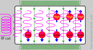
As you know, humans are mostly made out of water molecules and these molecules are found throughout our bodies in different ways. MRI machines can’t detect the electrons of hydrogen or the oxygen atoms; rather they “see” only the nucleus of the hydrogen atoms contained in the water molecule. These machines use properties of the hydrogen nuclei to produce MRI scans. As you might recall from honors chemistry, atoms have a property called “spin” and the spin of the hydrogen nuclei can be oriented in certain ways. The magnetic field of the MRI makes the spins of most of the hydrogen nuclei line up along the magnetic field. Few hydrogen nuclei however will have spins opposite to the magnetic field and these nuclei will be “high energy nuclei” while the others will have “low energy nuclei”. The green arrows in the picture below symbolize the magnetic field and the blue arrows show the spin.


When the low energy hydrogen nuclei are bombarded or “irritated” with energy, they respond in a way that allows us create a scan. But how does their response create a scan? Well every MRI machine has a coil of wire that produces enough energy to irritate the low energy hydrogen nuclei. When a current is applied to that coil, which is often called a RF coil, energy is produced in the form of a rapidly changing magnetic field, shown by the pink waves in the diagram.


This energy that is emitted  from the RF coil is the main building block of the MRI. The low energy nuclei absorb this RF energy and change their spin direction and become high energy nuclei.   


After some time, the RF energy is stopped and the recently turned “high energy nuclei” go back to their previous low energy state and as they do this, they start releasing the energy that was given to them. They release this energy in the form of waves and these waves are collected by “receiver coils” which is the blue coil in the animation. The receiver coils convert the energy waves into an electrical current signal and through this, the MRI machine can perceive the amount and concentration of hydrogen nuclei in the body.


Here you can see how an actual MRI scanner looks with all the pieces of the animations. 


While the physics behind an MRI is certainly confusing, I hope I cleared up some questions. I hope to get a more concise perceptive next week at my internship. Hope you all stick around till then!

Friday, February 12, 2016

First Day!


02/12/16

Hi everyone and welcome to my second blog post!


This week I didn’t get to spend too much time on-site at Mayo since my first official day was today, Friday. I focused some of my time this week on independent research. I attempted to decode several images Dr. Panda, my co-mentor, had sent to me prior to the start of my project to help with my proposal. I wasn’t able to decipher much at first, but now looking at it again after I went over the basics of a MRI machine today, it makes a bit more sense.


On my first day, Dr. Panda introduced me to many doctors including my other co-mentor Dr Silva. I toured the Radiology department in the Mayo Clinic Phoenix location and learned that they have seven MRI’s up and running almost all the time. We spent most of the time discussing how I would carry out my project and what they could do to assist. Most patients getting a MRI are either getting cranial scans or spinal scans but the patient scans that I will be looking at will be from the prostate and possibly the liver or pancreas. Dr. Panda and Dr. Silva explained to me that a MRI scan is composed of many series and a series is a picture that gives a different bit of information about what is being scanned. Each series comprises of different sharpness, resolution, etc. that tells us something new. Sometimes when people aren’t able to sit through a full MRI scan, doctors are left with only a few series or pictures and not the complete scan. Some series give more valuable information necessary for a diagnosis than others and with the help of Dr. Panda and Dr. Silva, I will be using a trial of prostate scans to determine which series are most important.

After deciding that using prostate scans will be most effective for the research project, Dr. Panda and I went to the Scottsdale campus of Mayo Clinic where I sat in on a meeting. I met with other doctors such as Dr. Min and Dr. James, both of whom I will be interacting with more later in my project. In the meeting they discussed, how to utilize the MRI machines so that there an optimal number of exams performed per hour. After the meeting, Dr. Panda and I returned to the Phoenix campus and finalized our schedule. Due to his and Dr. Silva’s unpredictable hours, the times when I will go in and shadow will be determined on a weekly basis. From my experiences today, I am looking forward to going in sometimes next week to meet with the rest of the team who I will be working with and to finally get started on collecting data for my project!


Friday, January 22, 2016

Who am I?

1/22/16

Welcome to my blog!


My name is Sparshee Naik and I am a senior at BASIS Scottsdale. For the last few months of my senior year, I will be doing a Senior Research Project called MRI: My Radiology Insight, where I will be spending my time at Mayo Clinic in the Radiology Department. I hope to keep you all updated on my experiences as I answer the research question in my proposal!


My Senior Research Project will be with Dr. Anshuman Panda, a radiologist at Mayo Clinic. I was offered an internship with him through another doctor, Dr. Amy Hara, who came to give a speech about professionalism at our school. When I asked her if she had any internship opportunities, she referred me to Dr. Panda and his research on the effectiveness of partial MRI’s vs. complete MRI’s. I am very excited to be working with Dr. Panda and as someone who comes from a family of doctors, I too want to be a physician in the future.


I became interested in medicine when I witnessed my grandfather perform a surgery at the young age of 10. I was amazed as he took a worm out of a woman’s eye, and it led me to the realization that thousands of processes are happening any given moment in our body to keep it functioning. My interest in medicine led to my attendance at a library program where students were shown the objectives of physicians; we used stethoscopes, saw ultrasounds, and even got to keep a copy of an X-ray of our hand.  I still have the X-ray to this day but seven years later, the X-ray makes as little sense now as it did then. My fascination with how to decipher information from that black and white picture still continues. I've always wanted to go into the medical field, and interning at Mayo Clinic will allow me to better understand the different fields. I am particularly interested in radiology because, whether it's in tv shows or real life, their diagnosis allows other doctors to assess a treatment; they carry a heavy authority. With Dr. Panda, I can further explore the daily life and responsibilities of a radiologist.

I will be meeting with Dr. Panda next week to discuss how often I can come in and when and also so I can get an overview of what my role will be as an intern. For ten weeks, starting February 4th, I will intern for at least fifteen hours a week at Mayo Clinic. It’s an experience I’m greatly looking forward to and I hope you stick with me as I start my internship!